Quantcast
Channel: cardiomyopathy – The American Journal of Medicine Blog
Viewing all 17 articles
Browse latest View live

Not What It Looks Like

$
0
0

Not What It Looks Like: A Transient Cardiomyopathy

12-735 Figure1_crop
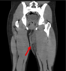
Significant cardiac signs and symptoms developed in a young patient undergoing treatment for a severe infection. The 23-year-old man presented with a left groin abscess and a 5-day history of fever. Computed tomography (CT) showed findings consistent with fasciitis (Figure 1). He was initially treated with intravenous vancomycin, ampicillin-sulbactam, and clindamycin. On hospital day 2, he developed severe chest pain, dyspnea, and nonsustained ventricular tachycardia.

The patient had a temperature of 99° F (37.2° C), a heart rate of 115 beats per minute, a blood pressure of 118/68 mm Hg, a respiratory rate of 35 breaths per minute, and an oxygen saturation of 95% on 100% oxygen delivered via a nonrebreather mask. Bilateral inspiratory crackles and a loud S3 gallop were present. A 12-lead electrocardiogram demonstrated sinus tachycardia with ST-segment depressions of 0.5-1 mm in leads V3 through V4 (Figure 2). An x-ray of the chest showed pulmonary edema and small bilateral pleural effusions (Figure 3). The patient’s troponin-T level was elevated at 1.12 ng/mL. Eight hours later, this peaked at 1.89 ng/mL. His creatine phosphokinase level was 726 IU/L; the creatine kinase-MB level, 73 ng/mL.

To read this article in its entirety, please visit our website. (If you follow the link to our website, you can view the videos submitted with this article.)

– Bryan J. Piccirillo, MD, Michael Gavin, MD, James D. Chang, MD

This article originally appeared in the June 2013 issue of The American Journal of Medicine.


Misconceptions and Facts About Hypertrophic Cardiomyopathy (video)

$
0
0

There are many misconceptions about cardiomyopathy. For example, it is thought that people with cardiomyopathy are destined to die young. In this interview, Dr. Joseph S. Alpert, editor-in-chief of The American Journal of Medicine, discusses a new review which outlines common misconceptions and facts about hypertrophic cardiomyopathy.

Related Research

Misconceptions and Facts About Hypertrophic Cardiomyopathy

Hypertrophic Cardiomyopathy as a Cause of Sudden Cardiac Death in the Young: A Meta-Analysis

$
0
0

sprinter on their mark ready to run

Sudden cardiac death is often linked with hypertrophic cardiomyopathy in young athletes, but with a divergence of study results. We performed a meta-analysis to compare the prevalence of sudden cardiac deaths associated with hypertrophic cardiomyopathy vs sudden cardiac deaths associated with structurally normal hearts.

Methods

A structured search of MEDLINE was conducted for studies published from 1990 through 2014. Retrospective cohort studies, patient registries, and autopsy series examining sudden cardiac death etiology in young individuals (age ≤35 years) were included. A random-effects model was applied to generate pooled summary estimates of the percentage of sudden cardiac deaths with structurally normal hearts at postmortem vs those caused by hypertrophic cardiomyopathy. Heterogeneity was assessed using I2. Subgroup analyses were conducted based on study location, patient age groups, and population types.

Results

Thirty-four studies were included, representing a combined sample of 4605 subjects. The overall pooled percentage of sudden cardiac deaths caused by hypertrophic cardiomyopathy was 10.3% (95% confidence interval [CI], 8.0%-12.6%; I2 = 87.2%), while sudden cardiac deaths with structurally normal hearts at death were more common (P <.001) at 26.7% (95% CI, 21.0%-32.3%; I2 = 95.3%). In nonathlete subjects, the pooled percentage of sudden cardiac deaths associated with structurally normal hearts (30.7%; 95% CI, 23.0%-38.4%; I2 = 96.3%) were significantly more common (P <.001) than sudden cardiac death caused by hypertrophic cardiomyopathy (7.8%; 95% CI, 5.8%-9.9%; I2 = 80.1%). Among athletes, there was no significant difference between summary estimates of hypertrophic cardiomyopathy and structurally normal hearts (P = .57), except in Europe where structurally normal hearts were more common (P = .01).

Conclusions

Hypertrophic cardiomyopathy is not a more common finding at death than structurally normal hearts in young subjects with sudden cardiac death. Increased attention should be directed toward identifying causes of death associated with a structurally normal heart in subjects with sudden cardiac death.

To read this article in its entirety please visit our website.

-Aditya J. Ullal, BA, Ramy S. Abdelfattah, MD, Euan A. Ashley, MRCP, DPhil, Victor F. Froelicher, MD

This article originally appeared in the May 2016 issue of The American Journal of Medicine.

Related Research

Power Failure: Acromegalic Cardiomyopathy

$
0
0
(A) The patient had bossing and macroglossia. (B) Swelling of the soft tissue, along with growth of the bone, cartilage, and muscle, produced enlargement of the hands. (C) The patient's shoe, size 19, is compared with a size 11 shoe.

(A) The patient had bossing and macroglossia. (B) Swelling of the soft tissue, along with growth of the bone, cartilage, and muscle, produced enlargement of the hands. (C) The patient’s shoe, size 19, is compared with a size 11 shoe.

Presentation

During his college years, the patient, at 6 ft, 8 in and 280 lb, aroused the attention of professional basketball scouts. But exertional fatigue and shortness of breath truncated his athletic aspirations. These symptoms continued in the ensuing years, necessitating approximately 15 hospitalizations at outside institutions for progressive dyspnea. No discernible etiology was established on any of these occasions. Now 35 years old, he presented to the Emergency Department reporting chronic shortness of breath, chest pain, and fatigue, symptoms of heart failure that had plagued him repeatedly in the past.

The patient was African American. He had no history of smoking, alcohol consumption, or illicit drug use. His family history was unremarkable. Because his medical care had been staggered, he was unable to provide definitive details about any of the therapeutic regimens he followed in the past or his compliance with them.

Assessment

Upon arrival, the patient was in moderate respiratory distress and was speaking in 3-word sentences. He was afebrile, his pulse rate was 85 beats per minute, his blood pressure was 193/111 mm Hg, his respiratory rate was 24 breaths per minute, and his oxygen saturation was 97% on 4 L of oxygen by nasal cannula. Physical examination disclosed frontal bossing, enlarged hands and feet, doughy skin texture, prominent skin tags, acanthosis nigricans around his neckline, and macroglossia. His jugular venous pressure was 12 cm, his lungs had diffuse crackles bilaterally, and the cardiac point of maximum impulse was laterally displaced. His cardiac rhythm was irregularly irregular; no murmurs, rubs, or gallops were evident, and 3+ pitting edema was noted below his knees, bilaterally.

Laboratory data were as follows: leukocytes, 4.3 × 103 cells/mm3; hemoglobin, 13 g/dL; glucose, 81 mg/dL; troponin I, 0.11 ng/mL (reference, <0.05 ng/mL); brain natriuretic peptide, 606 pg/mL (reference, ≤100 pg/mL). A chest film showed cardiomegaly, an enlarged right heart border, and pulmonary vascular congestion. Computed tomography of the chest demonstrated enlargement of all 4 cardiac chambers. Transthoracic echocardiography showed preserved biventricular function, severe concentric left ventricular hypertrophy (septum, 2.7 cm; posterior wall, 2.6 cm), and massive atrial enlargement (left, 12 × 16 cm; right, 12 × 6 cm). Cardiac catheterization identified elevated right and left heart pressures and mild narrowing of large-caliber (>4 mm in diameter) coronary arteries.

The findings pointed toward a diagnosis of acromegaly and associated cardiomyopathy. Further test results included growth hormone, 17.8 ng/mL (reference, 0.05-3 ng/mL), and for 2 separate samples, insulin-like growth factor-1, 700 and 771 ng/mL (reference, 81-225 ng/mL). A complete endocrine evaluation indicated that the patient had low testosterone and normal levels of luteinizing hormone, follicle-stimulating hormone, prolactin, and cortisol. Magnetic resonance imaging of the brain demonstrated a mild prominence at the left side of the pituitary gland, suggesting a 2-3-mm pituitary microadenoma. With additional questioning, the patient reported that his clothing and shoe sizes had increased over the previous decade; his shoe size went from 14 to 19, and he was unable to wear a cap on his head due to his enlarging forehead. This information further supported a diagnosis of acromegalic cardiomyopathy.


To read this article in its entirety please visit our website.

-Neha Maheshwari Mantri, MD, Ezra Amsterdam, MD, Marilyn Tan, MD, Gagan D. Singh, MD

This article originally appeared in the July 2016 issue of The American Journal of Medicine.

Demographics and Epidemiology of Sudden Deaths in Young Competitive Athletes (video)

$
0
0

sprinter on their mark ready to run


Sudden deaths in young, seemingly healthy competitive athletes are tragic events, often with wide media coverage and high public visibility. There is substantial interest in establishing the cause of these deaths and the roles that race and gender may play. In a new study published in The American Journal of Medicine, investigators report that more than one-third of recorded cardiovascular deaths were caused by hypertrophic cardiomyopathy, the majority in young male minority athletes. Sudden deaths due to genetic and/or congenital heart diseases are uncommon in females, but relatively common in African-American and other minorities compared to whites.

Researchers accessed the U.S. National Registry of Sudden Death in Athletes, 1980-2011 to define the epidemiology and causes of sudden deaths in competitive athletes. More than 2,400 deaths were identified in young athletes aged between 13 and 25 years engaged in 29 different sports. Over 840 athletes had cardiovascular diagnoses confirmed at autopsy.

“Utilizing this registry, we have established that hypertrophic cardiomyopathy is the leading cause of sudden cardiac death in male athletes and is an under-appreciated cause of sudden death in male African-American and minority athletes, but is a rare cause of death in female athletes,” explained lead investigator Barry J. Maron, MD, Tufts Medical Center, HCM Institute, Division of Cardiology, Boston, MA.

The investigators found that:

  • Male athletes were 6.5 times more likely to die from a sudden cardiac event than females.
  • More than one-third of deaths were caused by hypertrophic cardiomyopathy, which accounted for nearly 40% of male sudden deaths and was almost four times more common in males than females.
  • Cardiovascular death rate among African-Americans and other minorities exceeded whites almost five-fold.
  • Among cases of hypertrophic cardiomyopathy, more than 50% occurred in minority males, but only 1% in minority females.
  • Sudden deaths among male and female basketball players were three times more likely to be African-American and minorities than white.
  • Congenital coronary artery anomalies, arrhythmogenic right ventricular cardiomyopathy, and clinically diagnosed long-QT syndrome were more frequent among females.
  • Structurally normal hearts comprised less than 5% of athlete deaths.

Hypertrophic cardiomyopathy is a disease of the myocardium (heart muscle) in which a portion of the myocardium is hypertrophied (thickened) without any obvious cause, creating functional impairment of the cardiac muscle. It is a significant cause of sudden unexpected cardiac death in any age group and as a cause of disabling cardiac symptoms. There are often no symptoms beforehand.

“These observations underscore the potential value of the American Heart Association/American College of Cardiology recommended preparticipation screening in minority and other communities, particularly for the identification of hypertrophic cardiomyopathy,” Dr. Maron added.


Read the research:

Demographics and Epidemiology of Sudden Deaths in Young Athletes

$
0
0

running-stock


Sudden deaths in young competitive athletes are tragic events, with high public visibility. The importance of race and gender with respect to sport and the diagnosis and causes of sudden death in athletes has generated substantial interest.

Methods

The US National Registry of Sudden Death in Athletes, 1980-2011, was accessed to define the epidemiology and causes of sudden deaths in competitive athletes. A total of 2406 deaths were identified in young athletes aged 19 ± 6 years engaged in 29 diverse sports.

Results

Among the 842 athletes with autopsy-confirmed cardiovascular diagnoses, the incidence in males exceeded that in females by 6.5-fold (1:121; 691 vs 1:787,392 athlete-years; P ≤.001). Hypertrophic cardiomyopathy was the single most common cause of sudden death, occurring in 302 of 842 athletes (36%) and accounting for 39% of male sudden deaths, almost 4-fold more common than among females (11%; P ≤.001). More frequent among females were congenital coronary artery anomalies (33% vs 17% of males; P ≤.001), arrhythmogenic right ventricular cardiomyopathy (13% vs 4%; P = .002), and clinically diagnosed long QT syndrome (7% vs 1.5%; P≤.002). The cardiovascular death rate among African Americans/other minorities exceeded whites by almost 5-fold (1:12,778 vs 1:60; 746 athlete-years; P <.001), and hypertrophic cardiomyopathy was more common among African Americans/other minorities (42%) than in whites (31%; P ≤.001). Male and female basketball players were 3-fold more likely to be African American/other minorities than white.

Conclusions

Within this large forensic registry of competitive athletes, cardiovascular sudden deaths due to genetic and/or congenital heart diseases were uncommon in females and more common in African Americans/other minorities than in whites. Hypertrophic cardiomyopathy is an under-appreciated cause of sudden death in male minority athletes.

Sudden deaths in young competitive athletes are a highly visible medical and societal issue, which has attracted considerable interest in both the physician and lay communities.12345678910111213141516171819 Most recently, substantial attention has been directed toward the interaction of race, gender, cardiac diagnosis, and sport on sudden death risk, and how these variables may influence implementation of the most effective and practical strategy for preparticipation screening to detect unsuspected and potentially lethal genetic and/or congenital cardiovascular diseases.389101213141516171819 To that purpose, we have taken this opportunity to revisit these issues by accessing the largest available national forensic database, which represents a unique resource for insights into the epidemiology of sudden death events in young athletes and the underlying causes.


To read this article in its entirety please visit our website.

-Barry J. Maron, MD, Tammy S. Haas, RN, Aneesha Ahluwalia, Caleb J. Murphy, BS, Ross F. Garberich, MSc

To view additional an additional video relating to this story, follow this link.

This article originally appeared in the November 2016 issue of The American Journal of Medicine.

Achieving Extended Longevity and Quality of Life for Senior Patients With Hypertrophic Cardiomyopathy

$
0
0
Historical figures in hypertrophic cardiomyopathy. Left: Mr. Claude Brady, the first patient identified clinically with hypertrophic cardiomyopathy at age 22 years, is now 80 years of age. Right: Dr. Eugene Braunwald, who diagnosed Mr. Brady and went on to develop the first clinical profile of this disease in the early 1960s.

Historical figures in hypertrophic cardiomyopathy. Left: Mr. Claude Brady, the first patient identified clinically with hypertrophic cardiomyopathy at age 22 years, is now 80 years of age. Right: Dr. Eugene Braunwald, who diagnosed Mr. Brady and went on to develop the first clinical profile of this disease in the early 1960s.

To read this article in its entirety please visit our website.

-

This article originally appeared in the November 2017 issue of The American Journal of Medicine.

Elevated High-Density Lipoprotein Cholesterol Is Associated with Hyponatremia in Hypertensive Patients

$
0
0
Density plots comparing high-density lipoprotein cholesterol (HDL-C) at baseline in Systolic Blood Pressure Intervention Trial (SPRINT) participants who developed hyponatremia (red) during the study and patients who did not develop hyponatremia (blue). Baseline HDL-C was significantly more elevated in participants who later developed hyponatremia, for both males and females (P <.0001 in both sexes; Wilcoxon test).

Density plots comparing high-density lipoprotein cholesterol (HDL-C) at baseline in Systolic Blood Pressure Intervention Trial (SPRINT) participants who developed hyponatremia (red) during the study and patients who did not develop hyponatremia (blue). Baseline HDL-C was significantly more elevated in participants who later developed hyponatremia, for both males and females (P


Recently, the Systolic Blood Pressure Intervention Trial (SPRINT) showed that intensive lowering of systolic blood pressure is beneficial, but is associated with more adverse events. Hyponatremia was notably more frequent in the intensive treatment group. Investigating its risk factors is crucial for preventing this complication. Our objective in this study was to identify risk factors for hyponatremia in the adult population.

Methods

We investigated the baseline demographic, clinical, and laboratory data from the 9361 participants of SPRINT to identify the best predictors of hyponatremia (serum sodium ≤130 mEq/L), and adverse events, which could be attributed to hyponatremia, using machine learning and multivariable Cox proportional hazards models. We confirmed our results in the independent National Health and Nutrition Examination Survey (NHANES) cohort between the years 2005 and 2010 (16,501 participants).

Results

Elevated baseline high-density lipoprotein cholesterol (HDL-C) was a strong predictor of future hyponatremia. Multivariable Cox regression showed hyponatremia events to be significantly increased for SPRINT participants with baseline HDL-C levels in the highest quintile (hazard ratio [HR] 2.8; 95% confidence interval [CI], 2.2-3.7; P<.001), and were also associated with treatment-related serious adverse events (HR 1.6; 95% CI, 1.3-2.1; P<.001). We confirmed the association between HDL-C and hyponatremia in the NHANES cohort (HR 2.5; 95% CI, 1.7-3.7; P <.001).

Conclusions

Elevated HDL-C (≥62 mg/dL) is a risk factor for hyponatremia. Thus, hypertensive patients with elevated HDL-C should be closely monitored for hyponatremia when treated for hypertension.

To read this article in its entirety please visit our website.

-Ariel Israel, MD, PhD, Ehud Grossman, MD

This article originally appeared in the November 2017 issue of The American Journal of Medicine.


Importance of Genetic Testing in the Diagnosis of Transthyretin Cardiac Amyloidosis

$
0
0

doctor-patient-consultation-stock
To the Editor:

Systemic amyloidosis results from extracellular deposition of fibrillar material derived from aggregation of precursor proteins into insoluble beta-pleated sheets. The most frequently recognized types are due to prolonged inflammation, deposition of immunoglobulin light chains, and accumulation of transthyretin, a tetrameric protein synthesized in the liver. Transthyretin amyloidosis can occur owing to wild-type or autosomal-dominant mutant transthyretin and predominantly affects the heart and peripheral nervous system. We present here 2 cousins with rapidly progressive cardiomyopathy secondary to TTR p.Val40Ile, a rare variant with high penetrance.1 Before genetic testing, each was thought to have an alternative diagnosis.

In the first case, a 59-year-old Caucasian man with hypertension, coronary disease, and carpal tunnel syndrome presented with subacute dyspnea and mildly elevated troponin. Coronary angiography showed no obstructive disease; transthoracic echocardiogram showed severe left ventricular hypertrophy with ejection fraction 55% and moderate right ventricular dysfunction. Cardiac magnetic resonance imaging (MRI) demonstrated septal-predominant left ventricular hypertrophy with subendocardial late gadolinium enhancement in the left ventricle, septum, and right ventricular free wall. Serum amyloid A, free light chains, and urine/serum protein electrophoresis were unremarkable.

Endomyocardial biopsy was consistent with wild-type transthyretin amyloidosis by immunohistochemistry and liquid chromatography/mass spectrometry. However, he was referred for clinical trial enrollment, where genetic testing identified the mutation TTR p.Val40Ile. He underwent heart transplantation less than 2 years after his original diagnosis.

In the second case, a 58-year-old Caucasian man with hypertension and hyperlipidemia presented with dyspnea and was diagnosed with hypertrophic cardiomyopathy. Cardiac MRI demonstrated septal-predominant left ventricular hypertrophy with transmural and epicardial foci of late gadolinium enhancement. Ten years later, genetic testing was negative for hypertrophic cardiomyopathy-linked genes but heterozygous for TTR p.Val40Ile.

Transthoracic echocardiogram showed severe concentric left ventricular hypertrophy with ejection fraction of 20%-25%, right ventricular dysfunction, and thickened mitral leaflets with mild–moderate regurgitation. Endomyocardial biopsy confirmed amyloidosis; laboratory tests were without evidence of monoclonal gammopathy, and serum amyloid A levels were normal. He rapidly deteriorated and required heart transplantation less than 1 year later.

Amyloid diagnosis has historically relied on histologic demonstration of amyloid deposits in biopsy specimens, which reveal characteristic green birefringence under cross-polarized light after Congo red staining.2 It is vitally important to distinguish amyloid subtypes, given differences in management and need for family screening. Immunohistochemisty, although widely available, cannot identify the fibril type in 20%-25% of cases.3 Mass spectrometry has emerged as the gold standard for fibril typing, using computer algorithms to match peptides to a reference database. However, in one recent study of 56 genotype-positive transthyretin amyloidosis patients, liquid chromatography/mass spectrometry failed to identify pathogenic mutations in 9 individuals.4

Noninvasive cardiac imaging can assist in diagnosis and disease surveillance. Transthoracic echocardiography may demonstrate biventricular hypertrophy, valvular thickening, biatrial dilatation, and impaired strain.2 Bone tracer scintigraphy (eg, Tc99-pyrophosphate) is reported to be >99% sensitive and 86% specific for transthyretin amyloidosis,5 but is semiquantitative and does not evaluate cardiac structure or function. Cardiac MRI is emerging as an important diagnostic tool. In 263 patients with scintigraphic evidence of cardiac transthyretin amyloidosis, 79% had asymmetric septal hypertrophy, 71% transmural late gadolinium enhancement, and 96% right ventricular late gadolinium enhancement. Additionally, extracellular volume paralleled scintigraphic cardiac uptake and predicted mortality.6

Transthyretin amyloidosis is increasingly recognized in patients with heart failure, aortic stenosis, or a hypertrophic cardiomyopathy-like phenotype.7 Peripheral neuropathy, carpal tunnel syndrome, tendon tears, and spinal stenosis may also provide diagnostic clues to transthyretin amyloidosis.8 Although transthyretin amyloidosis treatment is currently limited, novel transthyretin-stabilizing and gene-silencing therapies hold promise for organ recovery and increased survival.2 A high index of suspicion and appropriate utilization of genetic testing can ensure timely and accurate diagnosis for patients and their family members.

To read this article in its entirety please visit our website.

-Jessica R. Golbus, MD, Joanna M. Wells, BS, Michael G. Dickinson, MD, Scott L. Hummel, MD, MS

This article originally appeared in the July issue  of The American Journal of Medicine.

Misconceptions and Facts About Hypertrophic Cardiomyopathy (video)

$
0
0
There are many misconceptions about cardiomyopathy. For example, it is thought that people with cardiomyopathy are destined to die young. In this interview, Dr. Joseph S. Alpert, editor-in-chief of The American Journal of Medicine, discusses a new review which outlines common misconceptions and facts about hypertrophic cardiomyopathy. Related Research Misconceptions and Facts About Hypertrophic Cardiomyopathy

Hypertrophic Cardiomyopathy as a Cause of Sudden Cardiac Death in the Young: A Meta-Analysis

$
0
0
Sudden cardiac death is often linked with hypertrophic cardiomyopathy in young athletes, but with a divergence of study results. We performed a meta-analysis to compare the prevalence of sudden cardiac deaths associated with hypertrophic cardiomyopathy vs sudden cardiac deaths associated with structurally normal hearts. Methods A structured search of MEDLINE was conducted for studies published […]

Power Failure: Acromegalic Cardiomyopathy

$
0
0
Presentation During his college years, the patient, at 6 ft, 8 in and 280 lb, aroused the attention of professional basketball scouts. But exertional fatigue and shortness of breath truncated his athletic aspirations. These symptoms continued in the ensuing years, necessitating approximately 15 hospitalizations at outside institutions for progressive dyspnea. No discernible etiology was established […]

Demographics and Epidemiology of Sudden Deaths in Young Competitive Athletes (video)

$
0
0
  Sudden deaths in young, seemingly healthy competitive athletes are tragic events, often with wide media coverage and high public visibility. There is substantial interest in establishing the cause of these deaths and the roles that race and gender may play. In a new study published in The American Journal of Medicine, investigators report that […]

Demographics and Epidemiology of Sudden Deaths in Young Athletes

$
0
0
  Sudden deaths in young competitive athletes are tragic events, with high public visibility. The importance of race and gender with respect to sport and the diagnosis and causes of sudden death in athletes has generated substantial interest. Methods The US National Registry of Sudden Death in Athletes, 1980-2011, was accessed to define the epidemiology […]

Achieving Extended Longevity and Quality of Life for Senior Patients With Hypertrophic Cardiomyopathy

$
0
0
To read this article in its entirety please visit our website. – This article originally appeared in the November 2017 issue of The American Journal of Medicine.

Elevated High-Density Lipoprotein Cholesterol Is Associated with Hyponatremia in Hypertensive Patients

$
0
0
  Recently, the Systolic Blood Pressure Intervention Trial (SPRINT) showed that intensive lowering of systolic blood pressure is beneficial, but is associated with more adverse events. Hyponatremia was notably more frequent in the intensive treatment group. Investigating its risk factors is crucial for preventing this complication. Our objective in this study was to identify risk […]

Importance of Genetic Testing in the Diagnosis of Transthyretin Cardiac Amyloidosis

$
0
0
To the Editor: Systemic amyloidosis results from extracellular deposition of fibrillar material derived from aggregation of precursor proteins into insoluble beta-pleated sheets. The most frequently recognized types are due to prolonged inflammation, deposition of immunoglobulin light chains, and accumulation of transthyretin, a tetrameric protein synthesized in the liver. Transthyretin amyloidosis can occur owing to wild-type […]
Viewing all 17 articles
Browse latest View live


Latest Images